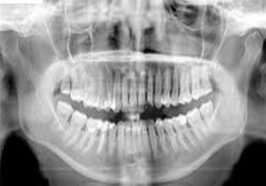
- It is a technique for producing a singletomographic image of facial structuresthat includes both maxillary andmandibular arches and their supportingstructures.
- Indications
• Trauma
• Location of third molars
• Extensive dental or osseous disease
• Known or suspected large lesions
• Tooth development
• Retained teeth or root tips
• TMJ pain • Dental anomalies etc
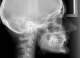
LATERAL CEPHALOMETRIC PROJECTION
- This view is used to evaluate facial growth and development, trauma, disease and developmental anomalies.
- This projection demonstrates the bones of the face, skull as well as the soft tissue profile of the face.
TRUE LATERAL SKULL
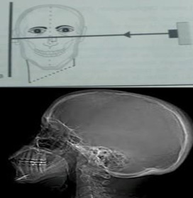
| The central ray is directed perpendicular to the cassette and the midsagittal plane and towards the external auditory meatus. |
- Fractures of the cranium and the cranial base
- Middle third facial fractures, to show possibledownward and backward displacement of themaxilla
- Investigation of the frontal, sphenoidal andmaxillary sinuses
- Conditions affecting the skull vault,
- Paget’s disease
- Multiple myeloma
- Hyperparathyroidism
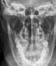
PA SKULL PROJECTION
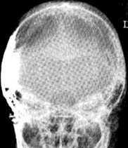
- The image receptor is placed in front of the patient, perpendicular to the mid sagittal plane and parallel to coronal plane,so that the canthomeatalline is perpendicular to the image receptor.
- Central Ray is directed at right angles to the film through the midsagittal plane through the occiput.
- to assess for medial and lateral displacements of skull fractures, in addition to neoplastic changes and Paget disease.
TOWNE’S VIEW
- The cassette is placed perpendicular to the floor.
- The long-axis of the cassette is positioned vertically.
- This is ananteroposterior view,with the back of the patient’s head touching the film. The canthomeatal line is perpendicular to the film.
- The central ray is directed at 30degrees to the canthomeatal line and passes through it at a point between the external auditorymeatus.
- It is primarily used to observe the occipital area of the skull.
- The necks of the condyloid process can also be viewed.
SUBMENTOVERTEX (BASE OF THE SKULL)
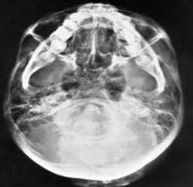
- The image receptor is positioned parallel to patient’stransverse plane and perpendicular to the midsagittal and coronal planes. To achieve this, the patients neck is extended as far backward as possible,with the canthomeatal line forming a 10 degree angle with the receptor.
- The central beam is perpendicular to the image eceptor, directed from below the mandible toward the vertex of the skull , and centered about 2cms anterior to a line connecting the right and left condyles.
- assessing potential pathology from trauma or disease progression to the basal skull structures, including the foramen ovale, foramen spinosum and sphenoid sinuses.
PA WATER’S VIEW (PNS)
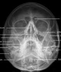
- The image receptor is placed in front of the patient and perpendicular to the midsagittal plane.
- The patients head is tilted upward so that the canthomeatal line forms a 37degrees angle with the image receptor.
- The central beam is perpendicular to the image receptor and centered in the area of maxillary sinuses
LATERAL OBLIQUE VIEW OF MANDIBLE
- The cassette is positioned against the patients cheek overlying the ascending ramus and the posterior aspect of the condyle of the mandible under investigation.
- The cassette is positioned so that its lower border is parallel with the inferior border of the mandible but lies at least 2 cm below it
- The positioning achieves a l0-degree angle of separation between the median sagittal plane and the film.
- The mandible is extended as far as possible.
- The centring position of the tube is the contralateral side of the mandible at a point 2cm below the inferior border in the region of the first/second permanent molar with angulation of 10 degrees cephalad or caudal
PA MANDIBLE
- The cassette is placed in front of the patient, so that the median sagittal plane should be perpendicular to the cassette. The head is then adjusted to bring the orbito-meatal baseline perpendicular to the cassette
- The cassette should be positioned such that the middle of cassette, is centred at the level of the angles of the mandible.
- The central ray is directed perpendicular to the cassette and centred in the midline at the levels of the angles of the mandible.
TMJ PANORAMIC VIEW
- The panoramic projection serves as the screening projection to identify odontogenic disorders and other disorders that may be the source of TMJ symptoms
- Gross osseous changes in the condyles may be identified such as asymmetries, extensive erosions, large osteophytes, tumors or fractures.
REVERSE TOWNE’S VIEW
- The image receptor is placed in front of the patient, perpendicular to the midsagittal and parallel to the coronal plane.
- The patient’s head is tilted downward so that the canthomeatal line forms a 25to 30 degree angle with the image receptor.
- To improve visualization of the condyles, the patient’s mouth is opened so that the condylar heads are located inferior to the articular eminence.
- The central beam is perpendicular to the image receptor and parallel to patient’s midsagittal planeand it is centered at the level of the condyles.
- Condylar neck and head
- High fractures of condylar neck, intracapsular fractures of the TMJ
- Condylar hypoplasia or hypertrophy
OTHER IMAGING MODALITIES
• CT • MRI • USG • CBCT